domingo, 24 de maio de 2015

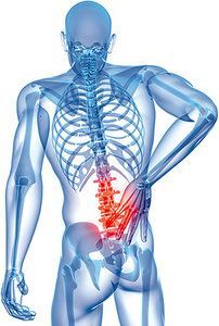
The Pain in the Low Back Often Begins in the Foot, or Is It in Your Head? What Do You Treat?


By Keith Innes
Ignoring the effects of the lower extremity on the functional capacity of the low back can mean missing the primary cause of the patients pain, and unnecessary treatment to another area. Following the kinetic chain to locate the pain generator is indeed a logical process, however it demands a significant amount of knowledge of the functional anatomy, arthrology and biomechanics (both normal and compensatory) of the lower limb and lumbopelvic regions, and a comprehensive grasp on the ascending and descending pathways of our nervous systems.
Chiropractic has always taken x-rays in a weightbearing position so that one can get an overall postural view of the patient, however the examination is for the most part a nonweightbearing situation, either supine or prone. This instantaneous "snapshot" of bony alignment may not reflect the functional positioning of the patient during propagation of the greatest forces causing deformities/malfunction during gait, or any other life-like situation. This is followed by an adjustment in yet another postural situation, most likely in an position which by no means correlates to or represents a real life situation.
The human biomechanical motion machine is not a static device, rather it is dynamic in every way and susceptible to gravity and ground reactive forces throughout our daily lives. To consider treatment of the low back in the absence of lower extremity examination and differential examination is illogical and unfair to our patients. Chiropractors have always suggested that we treat the cause. I totally agree, but what is really the cause? Always the spine? I do not think so!
Let me have you consider the following. In the journal of the lower extremity movement (Biomechanics, 2(9), Oct. 1995) there appeared an article, "Tracing the Pain." Do your patients complain of low back pain? Take a look at their legs and feet. Now you might be thinking that this is an insignificant article that just happened by chance. Let me assure you that it is not by chance, but by very exact design that others are looking to the cause of low back pain and finding the primary subluxation in the lower extremity. Remember what Dr. C. S. Gonstead stated: "Find it, accept it where you find it, fix it, and leave it alone." But you need to be competent to find it before the others are applicable. Diagnosing is just a matter of applying one's knowledge of anatomy, including neuroanatomy.
Gait is a very difficult concept to understand, but is very important to chiropractic. Gait is either gross gait or gait as it pertains to the individual joints. Gait from the Joint Point of View
At heel rocker/strike the calcaneus is inverted and the pre-tibial muscles are concentrically contracted. To keep the foot dorsiflexed the tibialis anterior muscle contracts just prior to heel strike. Recall that the this muscle attaches to the medial side of the first metatarsal, where it becomes intimately blended in with the fibers of the peroneus longus muscle. Together they form the longitudinal muscle-tendon-fascia sling which functions dynamically along with the inferior dropping fibula. The foot is lowered to the ground and is under the control of the eccentric contraction of the pre-tibial muscles. At this point the fibula drops inferior and loads the long head of the biceps femoris, the ipsilateral sacrotuberous ligament, the ipsilateral multifidus, the contralateral gluteus maximus, thoracolumbar fascia, and latissimus dorsi. Form and subsequent force closure of the iliosacral joint, on the side of stance leg, now initiates sacroiliac motion on an oblique axis. The tibia translates anterior in response to the body's line of forward progression.
Working in concert with concentric contraction of the pre-tibial muscles, the tibia translates over the talocrural joint. This position is known as midfoot rocker. The lower limb internally rotates throughout the contact phase causing the talus to internally rotate, plantar flex and adduct, thus initiating movement of the posterior portion of the subtalar joint. The subtalar joint is made up of the posterior, middle and anterior portions. The posterior has its own joint capsule and functions separately from the middle and anterior joints who share a common joint capsule with the navicular forming a functional joint -- the talocalcaneonavicular joint. The facet on the posterior calcaneus slides medially as the calcaneus moves to a position of a (right foot) counter clockwise motion in a sagittal plane. Simply put, the direction of slide of the articulation is opposite to the direction of movement of the calcaneus.
The middle and anterior portions of the subtalar joint move in the same direction as the calcaneus and, along with the navicular, plantar flex. The navicular also adducts at this point, as the midfoot and forefoot in the open kinetic chain position tends to follow the rear foot. This completion of subtalar pronation results in a simultaneous parallelism of the axes of the transverse tarsal joint to allow for pliant adaptation for accommodation to uneven terrain. This is known as mid foot pronation. The first ray complex (the first metatarsal and its associated cuneiform) follows the midfoot and pronates as well. This rotation reorients the first metatarsophalangeal joint axis obliquely, promoting toe pronation and the development of hallux valgus. Great toe pronation in hallux valgus is compensatory to eversion at the first metatarsocuneiform joint. Recalling that as the metatarsophalangeal joint pronates it also, due to the shape of its articulation, moves towards the midline of the now weightbearing forefoot. This results in a functional hallux valgus formation during the gait cycle. The tibia is between the necessary 10-20 degrees of anterior movement, and late mid-stance phase of gait is complete.
As the body moves anterior in a line of progression a number of events occur almost simultaneously: 
  • The contralateral hip moves forward, initiating internal or medial rotation of the femur; this in turn causes the stance phase leg to externally rotate.

  • The externally rotating leg causes the tibia, talus, and subtalar joints to supinate.

  • Ground reaction forces anterior to the metatarsal heads dorsiflexes the first toe, creating tension in the plantar aponeurosis. This pulls the great toe back, producing a reaction force at the first metatarsophalangeal joint, and a plantar flexion moment around the joints of the first ray complex.

  • Tension in the aponeurosis produces a supinatory movement around the subtalar joints, posterior and TCN.

  • The loaded metatarsal slides back over the sesamoids, assisted by contraction of the peroneus longus muscle which inserts into its base.

  • Eccentric contraction of the flexor digitorum longus helps to stabilize the first ray and medial arch.

  • The contraction of the peroneus longus, in addition to the above, also causes a convergence of the transverse tarsal joints, calcaneocuboid and talonavicular, which converts this articulation into a rigid lever for impending toe off.

  • Toe off is now complete and the procedure repeats itself.

In a previous article of mine, "The Back Force Transmission System," it was pointed out how critical the function of the lower limb was to the harmonious and dynamic functioning of the pelvis and spinal articulations.
The foot and gait are indeed complicated structures. To remain at the forefront of health care DCs need to understand this and much more. It should also be appreciated that higher centers (i.e., neocortex and thalamus to name but two) may predispose one to spinal and lower limb dysfunction. Information about these topics will follow in future columns.
The pain in the low back can start in the foot or it may be in your head. So what do you treat? Clearly from the above it can be seen that the case history and the examination, combined with a thorough knowledge of all aspects of anatomy and neurology, will lead you to the answer.
Keith Innes, DC
Ontario, Canada

Low Back Pain


The Appropriateness of Spinal Manipulation for Low Back Pain

By Paul Shekelle, MD, MPH
First I would like to tell the audience who I am and what RAND is and what role we play in this. I am an internist, and am on the faculty of UCLA. My research interests are in assessing the appropriateness and quality of health services, and I am the principal investigator of this project. 
The acronym RAND stands for research and development. RAND is a non-profit corporation performing research in the public good. RAND's research programs include classified defense research for the military, applied economics, education, sociology, civil justice, and health sciences. The health sciences department is the largest non-university-based research center in the U.S. For the past decade, a central effort of the health sciences program has been in the development and application of methods to assess the appropriateness of medical procedures.
As all of you in this room can appreciate, trying to assess the appropriateness of the use of any medical procedure is a difficult task. Ideally, we'd like to assess appropriateness on the basis of scientific evidence of increased benefit relative to risk. However, there are relatively few clinical problems that have been definitively answered by a well-executed controlled clinical trial. Even for these clinical problems which have been examined this way, frequently the patient in your office differs in some crucial way from the patients enrolled in the research trial such that generalizing the results from that trial to your patient may not be valid. And, of course, there are many clinical problems that have yet to be examined in a scientific way.
On the other hand, there is the large body of knowledge on appropriateness contained in the collective minds of the physicians of this country. Frequently, however, recommendations for action based on this clinical knowledge are in conflict, even among nationally recognized experts in the field. Resolving these conflicts can be problematic.
Lastly, there is the problem of the infinite variability of patients, making it an impossible task to try and define in advance the appropriateness of any medical procedure for any given patient that may come through your office door.
What can we hope to accomplish from studies of appropriateness? I believe we can define specific clinical circumstances for the appropriate use of medical procedures for general, commonly recurring classes of patients, that the clinician can use as a reference point, or guideline, to help him or her make the individual decision on appropriateness for the individual patient.
So how best to decide appropriateness? One way that has been developed, and I might add has been extensively used and is the method that has been the best studied, was developed at RAND and UCLA about 10 years ago, and has been undergoing improvement ever since. It involves a systematic literature review to capture all of the relevant literature; the development of clinically detailed indications describing patients who might undergo the procedure; the convening of a panel of experts; and the assessment of the indications by the experts for appropriateness, using a formal method designed to promote consensus, without forcing a lowest common denominator type of agreement. This method includes features of both the Delphi, namely that of anonymous ratings and multiple rounds of ratings with feedback of group results to individual members; and the Nominal group, namely the use of a roundtable, face-to-face discussion.
How did we use this method to assess the appropriateness of spinal manipulation for low back pain? First, we performed the literature review. We searched the Index Medicus and Medline for relevant articles, then searched the bibliographies of these articles for additional sources, and lastly had our sources reviewed by experts to ensure that we hadn't left out any critical material. This search netted 74 sources, which included 21 controlled trials of spinal manipulation as a treatment for low back pain. From these sources, a 32-page review was prepared, which summarized what is known about utilization, complications, and efficacy.
We then developed a detailed list of indications for patients who could potentially undergo spinal manipulation. This meant describing patients with back pain in a systematic fashion, in as much detail as possible, using clinically important variables, without using so many variables that we would overload our panelists. We did this by observing clinicians in their offices, and questioning them about their clinical reasoning. We spoke with experts, and reviewed the literature. We attempted to explicitly define key terms. The result was over 1,500 separate descriptions of patients with back pain, categorizing each patient by the length of the symptoms, the presence of comorbid diseases, the clinical course of the pain, the history and response to treatment of previous episodes of back pain, the findings on physical exam, the findings on lumbosacral radiographs, and the findings on CT or MRI if these tests were performed.
We then select a panel of experts. These are the general criteria governing the selection of RAND's expert panels. They have nine members; they are a mix of academic physicians and private practitioners; they include at least one member from each of the four major census regions of the country; and they include a mix of those who do the procedure and those who do not. This last point we feel is especially important. No medical specialty sees all of the patients with a given clinical problem. For instance, all patients with coronary artery disease don't end up in the cardiovascular surgeon's office; many patients are seen by cardiologists, internists, and family practitioners. The input from these practitioners is important, as they may see a different spectrum of the disease than the cardiovascular surgeon does. This is also true for back pain, as patients seek care from orthopedic surgeons, internists, GPs, and chiropractors, among others.
Given these considerations, this is the composition of the expert panel selected: three chiropractors, two of whom are in private practice and one of whom is an academic physician; two orthopedists and one osteopath, all of whom are distinguished experts in low back pain; one internist, who is an expert in back pain and is the principal investigator on the Back Pain Outcome Assessment Team; one family practitioner, who is trained in manipulation; and one person with degrees in many disciplines, a past president of the North American Spine Society, who practices as a neurologist.
We mailed the literature review, the definitions, and the 1,500 indications to the panelists. In private, they assigned an appropriateness rating, on a scale from one to nine, with one being highly inappropriate and nine being highly appropriate, to each of the 1,500 indications. They then mailed the completed appropriateness rating forms back to us, and we created summaries. All of the panelists met at RAND in Santa Monica. They were provided with the summary ratings, as well as a reminder of their own individual rating. We then had an open discussion of each indication for appropriateness, concentrating on areas of disagreement. This discussion was quite lively at times. After each general class of indications was discussed, each panelist privately rerated each indication for appropriateness. These second round ratings constitute the final results of the process.
What were the results? We define AGREEMENT as occurring when, after discarding the one highest and lowest rating, the ratings of the remaining seven panelists cluster within any three point range, that is two, three or four. We define DISAGREEMENT as occurring when, after discarding the one highest and lowest rating, at least one of the remaining seven panelists assigned a rating in the highest tertile (meaning most appropriate), and one of the panelists assigned a rating in the lowest tertile (meaning most inappropriate). By these definitions, on the initial round of ratings there was agreement on 12 percent of the indications and disagreement on 37 percent, with 51 percent indeterminate, meaning the panel neither agreed nor disagreed. After the final round, there was agreement on 36 percent of the indications, with disagreement on 12 percent. The number of indications changed somewhat between the two rounds, as the panelists altered the original indications to better fit clinical circumstances.
Comparing this panel to some previous RAND consensus panels. I find it interesting to note that this panel had the second lowest level of disagreement ever seen. For example, in a recently completed panel concerning the appropriateness of CABG and angioplasty, there was disagreement in the final round on 18 percent of the indications. The fact that a panel of orthopedists and chiropractors have less disagreement on the appropriate use of spinal manipulation for low back pain than a panel of cardiologists and cardiac surgeons have on the appropriate use of CABG and angioplasty is somewhat surprising.
From the nine point rating scale, we created three classes of appropriateness. We defined an indication as APPROPRIATE when after discarding the one highest and one lowest rating, the median of the remaining seven ratings was in the highest tertile, namely a seven, eight or nine, without disagreement. Analogously, we defined an indication as inappropriate when, after discarding the one highest and one lowest rating, the median of the remaining seven ratings was in the lowest tertile, that is a one, two or three without disagreement. Indications whose median rating was a four, five or six, and all indications with disagreement, were classified as equivocal, meaning they were neither clearly appropriate, nor clearly inappropriate.
Using these definitions, of the 1,550 final indications, 924 or 60 percent were classified as inappropriate, 112 or 7 percent were classified as appropriate, and 514 or 33 percent were classified as equivocal.
Which indications were appropriate, and which were inappropriate? Of course I can't go through 1,500 different indications at this time, but I can tell you the general characteristics of each type of indication.
In general, the appropriate indications tended to be those patients with acute low back pain, defined here as pain of less than three weeks; those patients with none or minor neurologic findings, minor neurologic findings being defined as either a unilateral diminished ankle reflex; no evidence of sciatic nerve root irritation, defined as typical radicular pain and the presence of a positive ipsilateral straight leg raising sign; physical findings of joint dysfunction, which in general meant hypomobility; unremarkable lumbosacral radiographs; and no adverse response to prior spinal manipulation, if any.
The general characteristics of those indications felt to be inappropriate were the following: no response or an unfavorable response to prior spinal manipulation; the presence of contraindications on lumbar x-rays, these being such things as malignant tumors, osteomyelitis, inflammatory arthritis, septic arthritis, acute or unhealed fracture, achondroplasia, and others; or no x-rays in the presence of risk factors for contraindications (such risk factors being fever, history of malignancy, severe osteoporosis, age greater than 50, significant trauma, and the like); pain of greater than six months duration, prior laminectomy (and this referred to manipulation within the area of prior laminectomy, not remote to it), major neurologic findings, these being the cauda equina syndrome, or progressive motor weakness; a lack of response to the current spinal manipulation; and the presence of a centrally herniated disc, spinal stenosis, or a large posterolateral herniated disc in the presence of sciatic nerve root irritation or any neurologic findings.
In the context of this discussion on the continued appropriateness of spinal manipulation in the face of a lack of response to current spinal manipulation, there was unanimous agreement on the following: An appropriate trial of spinal manipulation for patients with low back pain is two weeks of treatment for each of two different types of manipulation, after which, in the absence of improvement, continued manipulation is not indicated.
What were the characteristics of equivocal indications? Sciatic nerve irritation is the clinical factor responsible for the greatest number of equivocal indications with disagreement. The presence of a herniated disc on CT/MRI, especially in the absence of signs or symptoms other than back pain, was also a characteristic of many equivocal indications. The longer the patient had back pain, the more likely the indication was to be equivocal.
It is important to keep in mind that the percent of indications given a rating of "appropriate" does not translate into the percent of patients with back pain who might be appropriate for spinal manipulation. Patients with the cauda equina syndrome, or malignancies, or an abdominal aortic aneurysm, or who take coumadin are all felt to be inappropriate candidates for manipulation, while patients with uncomplicated acute low back pain are felt to be appropriate candidates. In terms of numbers, the inappropriate indications I just listed outnumber the appropriate indications by four to one, but my guess is that the average chiropractor sees many more patients with uncomplicated acute low back pain than patients with the cauda equina syndrome. We don't accurately know what kinds of patients are presenting to clinicians' offices and subsequently undergoing spinal manipulations. Future research is needed to answer this question.
Several important limitations deserve mention. The first is that we dealt only with spinal manipulation, and not the spectrum of conservative medical or chiropractic care. Such care may include other forms of treatment that may prove beneficial for patients with back pain. Secondly, the alternative treatment against which spinal manipulation was being compared was not explicitly defined. Rather, each panelist was comparing spinal manipulation against his own internalized "best available alternative," and these alternatives may have differed between panelists. One panelist may have been comparing spinal manipulation with bedrest and analgesics, while another, for the same indication, may have been comparing it with surgery. In the future, we will need to be more explicit about alternative treatments. Lastly, patient preferences are not accounted for in this process. Such preferences can greatly affect the appropriateness of procedures. Some patients may prefer a trial of spinal manipulation rather than bedrest, while others may prefer the reverse.
To summarize, let me state that this conference was not the "final word" on the appropriate use of spinal manipulation for low back pain. Rather, it is a first step, in that for the first time, orthopedists, chiropractors, and others assessed the appropriateness of spinal manipulation using a well-established method developed for such assessment. Our results show that there are significant areas of agreement, for both appropriate and inappropriate indications. Additionally, areas of disagreement are clearly defined. Finally, a number of indications for which the panel felt that there was not enough scientific data or clinical experience to assess appropriateness have been identified, and these areas may be fruitful avenues for future research.
Paul Shekelle, M.D., M.P.H.
The RAND Corporation
Santa Monica, California

Low Back Pain


Rehabilitation of Low Back Pain Patients

By Alan Jordan and Keld Ostergaard, DC, MD
About the authors: Dr. Keld Ostergaard, a 1990 graduate of the Anglo-European Chiropractic College, has been in private practice since 1991. He received his MD from the University of Copenhagen (1995), and that institution's gold medal for research (1994) in molecular biology and osteoarthritis. 
He is co-editor of the journal of the Danish Chiropractic Assoc., and an FCER research fellow. His articles on rehabilitation and assessment of the low back and neck have been published in the Journal of Musculoskeletal Pain, in the journal, Pain, and in Spine (June, 1995 editorial). The Jan. 1996 issue of JMPT featured his double article on neck/shoulder rehabilitation. Dr. Ostergaard is pursuing his doctorate at the University of Copenhagen.
Dr. Alan Jordan is a 1979 graduate of the Canadian Memorial Chiropractic College, and has been in private practice since that time. He is in his final year of a doctoral program at the University of Copenhagen's National University Hospital. He is a research fellow of the FCER and the Danish Chiropractic Assoc., and has been a consultant to Denmark's Dept. of Health.

In our first article regarding the topic of rehabilitation in chiropractic clinics (Jan. 1, 1995, "DC"), we reviewed what we felt were the most compelling reasons for chiropractors to incorporate rehabilitation in their overall treatment strategies for patients suffering both chronic and recurrent spinal pain. We presented some of the international developments in rehabilitation and emphasized that excellent clinical results could be obtained without employing high-tech and prohibitably expensive equipment. In this article we will discuss the essential aspects of program design for low back rehabilitative protocols.
Trends in Low Back Rehabilitation
Patients suffering from low back pain have received advice regarding exercise programs from health care workers for hundreds of years. The design of these programs has been largely based upon empirical considerations, and only in the recent past have researchers begun to scrutinize the validity of exercise for low back pain and program design.1 Research studies have clearly demonstrated that the clinical results obtained from exercise programs undertaken in clinics and under constant supervision are clearly superior to those programs carried out outside of the clinical setting.2 Patient compliance is extremely poor when subjects are not monitored. Effective training requires equipment so that the elementary principles of rehabilitation including progressive resistance can be adhered to while patient safety is maintained. Patients who have suffered low back pain for longer periods of time need encouragement and guidance from instructors and they feel secure training at the clinics where knowledge of their particular case is greatest.
During the '70s and early '80s, there was a theoretical debate going on between adherents of William's protocol, which emphasized strengthening of the abdominal musculature and stretching of the posterior musculature, and proponents of McKenzie's protocol, which emphasized training the posterior musculature of the spine. It is beyond the scope of this paper to review the debate, but as with so much else in physical medicine it needed to be put to the test. Two studies involving both acute and chronic patients clearly demonstrated that strengthening the extensors of the spine as opposed to the abdominal musculature resulted in superior results.3,4 Since that time the clinical studies of Claus Manniche MD, PhD and others, have clearly demonstrated that excellent results can be obtained on both chronic and postoperative patients by strengthening primarily the posterior musculature.1
A recent study has shown that manipulation combined with extension exercises resulted in superior results to those obtained through exercise alone.5 Functional assessment of patient populations compared with matched healthy individuals repeatedly points to decreased strength levels of the posterior musculature, resulting in patients which are "flexor powered."6 In the healthy population the extension to flexion strength ratio is approximately 1.5 to 1. Patient populations demonstrate one to one extension to flexion ratios, thus indicating that the greatest strength losses occur in the posterior musculature.6 Epidemiological studies have also shown that reduced endurance levels of the extensors predispose patients to repeated episodes of low back pain,7 as well as healthy individuals involved in heavy work tasks who have not experienced previous low back pain.8
Although research authorities recommend strengthening both the extensors and the abdominal musculature,1 clearly the emphasis must be placed on strengthening the extensor musculature.
Another aspect of exercise that has resulted in debate among health professionals is the type of exercise patients should be doing. Here the debate has revolved around whether patients should be doing their exercises isometrically, isotonically, isokinetically, and so forth. After Nachemsom and Elfstrom published their disc pressure measurements in the early '70s,9health workers reverted to isometric exercise to decrease the possible increases in discal pressure which could result from dynamic exercise. A few studies published in the '60s resulted in positive results,10,11 but clearly the trend today is towards full range of motion dynamic exercise.1 This provides increased blood perfusion to the muscles and increases strength throughout the range of motion trained. Additionally, the many published studies utilizing dynamic exercise report exceedingly few negative side-effects from dynamic exercise. At the present time there is no evidence that superior results can be obtained through predetermined speeds during training, such as those employed in isokinetic training.
Designing a Low Back Exercise Program
Exercise programs can be divided into three major phases.
The first is the warm up phase, requiring 10-15 minutes. Here patients increase body temperature by stationary bicycling or on step machines. This should be carried out for 6-8 minutes. Increased body temperature increases tissue elasticity and blood is channeled to the musculature. Afterwards all relevant muscles should be stretched in preparation for strength training.
The second phase involves strengthening of the muscles which the chiropractor has determined necessary. In this article we will concentrate on the extensors of the spine, as we feel that they are the most important muscle group requiring rehabilitation. This period usually lasts 35-45 minutes. The extensors can be strengthened in both the sitting position or in the prone position. Exercising the extensors of the spine in the sitting position is usually carried out in specially designed training machines with resistance and isolation of the extensor muscles. The resistance may be variable and patients are generally seated in fixed positions.
image - Copyright – Stock Photo / Register Mark
Another way to strengthen the extensors is in the prone position (Picture 1). This has the advantage of improving the coordination of the different muscle groups involved in lifting. The first muscles to be activated are the hamstrings, followed by the gluteals, and finally the extensors of the spine. On the way down the opposite sequence takes place. This motion duplicates placing an object on the floor and thus provides another valuable coordination improvement. For most patients the resistance level with this type of exercise is roughly 30-35 percent of their maximal capabilities. This is the ideal level for endurance training and provides for maximal blood perfusion and healing. Progressive resistance can be provided by asking the patients to carry weights in their arms or by providing them with vests which contain weights. Patients begin at approximately 30 to 40 repetitions in series of 10, resting for 1-2 minutes between series, and gradually increasing to approximately 100 repetitions during a three month period.
Patients who have difficulty carrying out an extension can be assisted by an instructor until they can manage themselves. This type of training is ideal to overcome fear avoidance, but should not be continued for more that a few weeks.
image - Copyright – Stock Photo / Register Mark
Hyperextension (Picture 2) has been the subject of heated debate. After having trained hundreds of subjects and having conferred with others in rehabilitation we feel that there is not much to be gained by hyperextending much more that 20-25 degrees over the horizontal. One need not be hysterical however about extending over the horizontal because to strengthen the segmental musculature some extension over zero degrees is necessary. In a future article regarding the effect of training on different tissue types we will expand on hyperextension and its particular effect on the discs of the spine.
Other muscles requiring training include the gluteals, the quadriceps and the abdominals. Fitness training may be integrated but is not a necessity.
The concluding phase of each training session should include relaxation and stretching of the trained muscles. This cool-down phase should last for approximately 10 minutes making the entire session somewhere between one hour and 15 minutes to one hour and 30 minutes.
Most chronic patients will require 2-3 training sessions per week for a period of at least three months. Some patients will require more time depending upon the duration and severity of symptoms and progress during rehabilitative procedures. It is not unusual for rehabilitation to require upwards of six months to maximize both objective and subjective gains.
The majority of chronic low back patients, or those suffering recurring pain, require rehabilitation. The most logical place for rehabilitation is in the clinical setting.
In future articles we will discuss among other topics contraindications and possible side-effects to rehabilitation, patient selection and expectations, monitoring results and other relevant aspects of rehabilitation in the chiropractic clinical setting.
References 
  1. Manniche C. Assessment and exercise in low back pain. Doctoral thesis. Laeforenings Forlag. Copenhagen 1995.

  2. Reilly K, Lovejoy B, Williams R, Roth H. Differences between a supervised and independent strength and conditioning program with chronic low back syndromes. J Occup Med 1989; 31: 547-50.

  3. Nwuga G, Nwuga V. Relative therapeutic efficacy of the Williams and McKenzie protocols in back pain management. Physio Prac 1985; 99-103.

  4. Donelson R, Grant W, Kamps C, Medcalf R. Pain response to sagittal end range spinal motion: A prospective, randomized, multicentered trial. Spine 1991; 16: 206-12.

  5. Erhard RE, Delitto A, Cibulka M. Relative effectiveness of an extension program and a combined program of manipulation and flexion and extension exercises in patients with acute low back syndrome. Physical Therapy 1994; 74: 1093-99.

  6. Beimborn DS. Morrisey MC. A review of the literature related to trunk performance. Spine 1988; 13: 655-60.

  7. Biering-Sorensen F. Physical measurements as risk indicators for low-back trouble over a one-year period. Spine 1984; 9: 106-19.

  8. Luoto S, Heliovaara M, Hurri H, Alantra H. Static endurance and the risk of low-back pain. Clinical Biomechanics 1995; 10: 323-324.

  9. Nachemson A, Elfstrom G. Intravital dynamic pressure measurement in lumbar discs. Scandinavian J of Rehab Med. 1970; suppl 1: 1-40.

  10. Kendall Ph, Jenkins JM. Exercises for backache: A double-blind controlled trial. Physiotherapy 1968; 54: 154-7.

  11. Lindstrom A, Zachrisson M. Physical therapy on low back pain and sciatica. Scand J Rehab Med 1970; 2: 37-42.

Alan Jordan, DC
Keld Ostergaard, DC, MD
Farum, Denmark

Low Back Pain


 Low Back Painand Episacral Lipomas

By David Bond
A cause of low back pain and disability often overlooked by practitioners who treat patients suffering from acute and chronic musculoskeletal complaints is the episacral lipoma. Although usually thought of as a minor condition, it is capable of producing considerable low back pain.
First described by Ries in 1937,1 episacral lipomas are small, tender, "tumor-like" nodules occurring mainly over the sacroiliac region which can cause disabling low back pain. The term "lipoma" is descriptive only in that through direct palpation over the region, the examiner is able to detect a subcutaneous mass similar to the benign tumors; however, it is not a tumor, but is subfascial fat which has herniated through the overlying fascial layer.
Perhaps a better term is that of the lumbar fat herniation as described in 1944 by Copeman and Ackerman.2 In their research, they reported 10 cases of severe and disabling low back pain in which they identified the fatty tumors as the principle cause of the patient's complaints. They subsequently excised the herniation, which produced striking relief of the pain. In 1945, Hertz reported the cases of six women with excruciating low back pain.3 All of the women had a history of a traumatic strain prior to its occurrence of, which was sometimes accompanied by unilateral leg pain. The low back pain in all six women was dramatically relieved by the removal of a herniated fat tumor.
In a followup study by Copeman and Ackerman, 11 new cases were described.4 In all 11 patients, a biopsy confirmed the presence of edematous fat lobules herniating through deficiencies of the fibrous compartments. It was felt that the pain was produced in the fibro-fatty tissue and not in the musculature itself.
Hucherson and Gandy reported in 1948 that of 32 patients who had undergone surgical removal of the lipoma, only two patients failed to experience relief of pain.5 Many other researchers have reported that in patients with backaches and occurrence of the nodules, relief was obtained immediately by injection of a local anesthetic and some by operation. At times, dramatic relief was obtained, and there was no recurrence of pain over time.6,7,8,9 In a study by Singewald,10 1,000 persons were evaluated for lipomas. They were found in 16% of the subjects; however, only 10% had reported back pain. Therefore, it is not an uncommon finding in the general population, although it is usually asymptomatic.
Fat herniations occur in predictable sites along the edge of the sacrospinalis muscle just above the iliac crest, very close to the natural "dimple" in the sacroiliac area.10 In this area, through abnormal tension, trauma, or by inherent weaknesses of the fascia, as well as through foramina for cutaneous nerves, the underlying fat pad may herniate through the fibrous tissue between the superficial and deep layers. Copeman and Ackerman4 mapped the basic fat pattern of the lumbar region from 14 cadaver studies with reference to the most common sites for the occurrence of fat herniations, which were felt to be extremely corresponding. The researchers reported that during dissections, it was not uncommon to find the fascia to be of non-uniform thickness. They also found actual deficiencies of the fascia in which underlying fat tended to bulge through.
In addition, they were able to describe three basic types of herniations: pedunculated, nonpedunculated and foraminal. The nonpedunculated hernia appears as a tense swollen nodule, which protrudes frequently along the iliac crest. Pedunculated hernias have the appearance of a strangulated polyp through the fascia connected by a fibrous pedicle. In the foraminal type, the fat herniates through the foramina containing the cutaneous branches of the posterior rami of the first three lumbar nerves as they pierce the deep fascia after leaving the body of the muscle. A horizontal fold of membrane acts as a valve that prevents the herniation from occurring during flexion of the back; however, a failure to function normally may result in a herniation. Of the three, the nonpedunculated herniation appears to be the most common. Biopsy of the specimens revealed that they were composed entirely of normal adipose tissue with some edema present. In some cases, there was evidence of patches of fibrous tissue growing in the fatty tissue and others with nerve tissue present; however, this has not been a consistent finding. From a clinical standpoint, the mechanism of pain is not fully understood; however, pain appears to be the primary feature, which seems to be due to the expansion of the fat herniation in the otherwise unyielding fibrous capsule, in that removal of the lipoma alleviates the pain.
The pain pattern of the fat herniation originates in a focal region; however, it may radiate in an ill-defined distribution and may be variable in intensity and duration.9 Upon palpation, the patient is usually able to describe the exact point of extreme, or pinpoint, tenderness. It is different from a trigger point as described by Travell13 in that the examiner can palpate a definite mass rather than a taut band of skeletal muscle. However, like a myofascial trigger point, firm pressure may produce pain that radiates in a general and segmental distribution.11
Depending upon the severity of the pain, there may be a restriction of the lumbar range of motion, and the pain may increase with positioning.9 There may well be a significant degree of paraspinal muscle spasming which may also be related to the referred pain, as well as the nature of the original incident.2 No specific structural abnormality of the spine has been identified. Nerve root traction tests are usually normal, with a production of primarily low back and sacral pain upon testing, unless there is a concomitant disc herniation.8 Reports of pain radiating down the side affected with the lipoma are frequent; however, there is no uniformity of the radiation area.5 Diagnosis is usually confirmed by the injection of local anesthetic, which significantly alleviates the pain, at least temporarily.12
The posterior elements of the lumbar spine are innervated by branches of the lumbar dorsal rami separate from conditions produced by the intervetebral disc, referred pain or syndromes mediated by other nerves.14 Any structure innervated by the lumbar dorsal rami is potentially capable of being a primary source of mechanical pain, including the zygapophyseal joints, ligaments, muscles and their fascia. Included in these possible sources are: strains; disc disease; degenerative arthritis; facet syndrome; spondylosis; spondylolisthesis; and subluxations. Psychogenic and iatrogenic disorders must be further ruled out.
It is reasonable to assume that due to the nature of the pain, heat, massage and manipulation would be the treatment of choice, with steroid injections reserved for primarily diagnostic purposes, and surgery as last resort. The typical patient as described by Singewald, however, presents with unilateral low back pain, some radiation of pain to the buttock or thigh, and a fairly long history of symptomatology. Furthermore, having had evaluations by medical, chiropractic, acupuncture, gynecological, and other health specialists, and with negative x-rays or other diagnostic studies, the patient finds no relief from the traditional forms of physiotherapy. No studies from a purely chiropractic standpoint have been submitted as to whether or not manipulation of the pelvis, sacrum or vertebrae provide any significant relief.
Current Case Study
A 39-year-old male was referred to the clinic for evaluation of a work-related injury to his low back. His presenting complaints were low back pain radiating to the left lower extremity. He had previously been seen by a chiropractor but had not responded to manipulation. Due to signs of neurotraction, an MRI was obtained, revealing multilevel disc bulges with obscuration of the left L5-Sl nerve root. Nerve conduction and EMG testing were then performed, revealing chronic denervation of the left L5-Sl nerve root.
The patient was referred to an orthopedic surgeon and subsequently underwent lumbar laminectomy and directory. Following surgery, as well as a prolonged course of physical therapy and exercise rehabilitation including ultrasound, iontophoresis and EMS, the patient stated that overall, his leg pain had resolved; however, he continued to experience low back pain which was aggravated by lying supine, as well as flexion and bending. He approximated that 30% of his pain remained in his low back. When asked to point to the greatest area of pain, he pointed to the left episacral region with radiation to the hip.
Palpation revealed a hard nodule of approximately two centimeters in diameter directly overlying the PSIS. Firm pressure reproduced complaints of low back pain. A second nodule was palpated on the opposite side; however, this was asymptomatic. He was then prepped for injection of a mixture of dexamethasone and zylocaine. Upon injection, he experienced immediate relief of the low back lasting between 2-3 hours. In addition, he was able to forward-flex at the waist without difficulty. Subsequent injections also alleviated pain for 2-3 hours, but no permanent relief was obtained. Due to his postsurgical status, no forceful spinal manipulation was attempted.
Although absent from most differential diagnoses, which have a tendency to show a strong preoccupation towards the discs and vertebrae, the episacral lipoma is fairly common, with symptomatology similar to other conditions. As this case shows, the episacral lipoma may account for a significant degree of pain in patients with mild disc bulges, even in those patients who require low back surgery, and may account for the prolonged pain symptomatology following surgery.
It is apparent that residual pain from the lipoma may be aggravated by prolonged bedrest and certain movements. For doctors of chiropractic, the episacral lipoma may be significant in of the general population. Are these patients the ones who complain of intractable low back, and have tried the gamut of conventional medical treatment, only to miraculously respond to manipulation; or are these patients considered the chiropractic failures in that manipulation will not repair a fascial tear or reduce a herniated lipoma? Is it possible that sacroiliac joint dysfunction, pelvic unleveling or vertebral subluxation is somehow predisposing to the herniation, due to altered biomechanics, which may add more tension to the fascia?
To date, research in the chiropractic profession is lacking, and no published studies have been recorded. According to published medical studies, the treatment of choice is injection, with excision of the lipoma and repair of the fascial wound occurring in severe cases. The cases and history have been thoroughly duplicated many times from a medical standpoint.
The episacral lipoma should be considered more often in the workup and differential diagnosis of acute and chronic low back pain, especially in instances of work-related injuries owing to the traumatic nature of the herniation. Furthermore, it is a condition in which doctors of chiropractic and medical doctors may have much to gain for their patients for inter-referrals.
References 
  1. Ries E. Episacroiliac lipoma. Amer J Obstet Gynec 1937;34:490.

  2. Copeman WSC, Ackerman WL. "Fibrositis" of the back. Quart J Med April-July 1944;13:37-51.

  3. Herz R. Herniation of fascial fat as a cause of low back pain. JAMA 1945;128:921-925.

  4. Copeman WSC, Ackerman WL. Edema or herniations of fat lobules as a cause of lumbar and gluteal "fibrositis." Arch Int Med 1947;79:22.

  5. Hucherson DC, Gandy JR. Herniation of fascial fat. Am J Surg 1948;76:605-609.

  6. Dittrich RJ. Coccygodynia as referred pain. Am J of Bone and Jt Surg 1951;33-A:715-718.

  7. Bonner CD, Kasdon SC. Herniation of fat through lumbodorsal fascia as a cause of low-back pain. New Eng J of Med 1954;251:1102-1104.

  8. Dittrich RJ. Soft tissue lesions as a cause of low back pain. Am J of Surg 1956;91:80-85.

  9. Wollgast GF, Afeman CE. Sacroiliac (episacral) lipomas. Arch Surg 1961;83:925-927.

  10. Sinqewald M. Sacroiliac lipomata: an often unrecognized cause of low back pain. Bull Hopkins Hosp 1966;118:492-498.

  11. Pace JB. Episacroiliac lipoma. Am Fam Phys 1972;6:70-73.

  12. Faille RJ, Low back pain and lumbar fat herniation. Am Surg 1978;44:359-361.

  13. Travell JG, Simons DG. Myofascial Pain and Dysfunction. Baltimore: Williams & Wilkins, 1985.

  14. Bogduk N. Lumbar dorsal ramus syndromes. Chapter 38. In: Greive GP (ed.) Modern Manual Therapy of the Vertebral Column. New York: Churchill Livingston, 1986.

David Bond,DC
Encino, California

Exercises for Back Pain


Exercises for Back Pain: Low-Compression Training Program

By Joseph D. Kurnik, DC
Editor's note: While technically not a part 2, this article expands upon concepts introduced by Dr. Kurnik last year in "Low Compression Resistance Exercises for Back and Neck Pain Sufferers" (Nov. 4, 2012 issue).
This program is intended for two groups of people: 1) those who want to engage in resistance exercises for the major regions of their body without developing back pain in the process; and 2) those who already have back pain and want to do resistance exercises, but consistently re-irritate their back when trying to do so. There is a point at which traditional methods of exercise fail to do what is hoped for: a more pain-free and fit body with increased endurance and muscle tone. This does not make traditional training methods bad or wrong. This simply means that for many, a new and safer path is needed at a certain point.
In general, what we are dealing with are the effects of two forces, compression and decompression. Compression is going on in the spine every day, every moment we sit or stand. The weight of the body and gravity cause compressive effects on the intervertebral discs andfacet joints. The disc bears about 80 percent of the spinal compressive load, and the facets bear the remaining 20 percent.
The natural way to decompress the spine, of course, is to lie down. Pressures are lowest in the spine when lying down and highest when sitting and standing. Bending, slumping, twisting, reaching and lifting things (such as weights) will significantly raise the pressures in the discs and facet joints. Lying down, doing inversion by hanging, doing certain exercises and spinal traction will lower disc pressures.
There is actually a battle going on each day in the spine between compression and decompression. The effect of compression is to squeeze the disc, forcing water out of it and gradually changing its composition. The other effect is the consequential thinning of the disc as it loses water. As the disc thins, it puts more weight on the facet joints and they become arthritic. The net result is that the holes through which nerves exit the spine become smaller and can cause pinching of spinal nerves. This can cause leg pain or sciatica.
As the disc degenerates during thinning, its outer fibers can become damaged enough to contribute to back pain. Pressure on the facet joints can cause back and leg pain. This is all attributed to compression as a major element, combined with other simultaneous body movements, such as bending and twisting. Add weights to that, and you accelerate the effects of compression.
The goal is to reduce compression as much as possible, especially while performing weighted exercises, and to add elements of decompression as much as possible.
Cautions / Considerations
In order to avoid or minimize spinal pressures during resistance and compression exercises, certain principles need to be followed. In general, avoid standing and seated exercises using free weights. Avoid using machines in the upright position that exert vertical pressure on the spine, such as standing squats or seated presses above the shoulders. We are trying to reduce the vertical compressive loads on the spine. Other considerations are as follows:
  • Avoid standing extension and flexion exercises. These increase vertical spinal compression.
  • If sciatic pain or numbness is present, avoid hamstring stretching.
  • Avoid sitting or standing exercises whereby the head is put into extension, i.e., bending backward.
  • Avoid exercises that require sitting / standing and raising the arms higher than shoulder level (exceptions include cable bar pulldowns for the back). You don't want to do presses above the head or shoulders.
  • Avoid picking up free weights when standing or sitting.
  • Avoid hard-floor-impact exercises, jumping or twisting.
  • When lying on your back, keep the knees up and feet flat. This reduces the lumbar curve and spinal pressure.
  • Avoid ball exercises. Trying to keep oneself balanced causes stressful positioning and tensing of muscles. Tensed muscles contract, shorten and compress the spine.
  • Avoid lunges if back arching is painful. Do not hold weights while lunging.
In order to give more perspective, consider an experiment conducted in Sweden [Nachemson AL. "The Lumbar Spine, an Orthopaedic Challenge." Spine, 1976;1(1):61], that demonstrated relative spinal pressure in lower lumbar discs in various positions and with various exercises. These are listed below with their relative values. They are relative values, but for illustration, think pounds per square inch in the lower lumbar spine.
  • Standing: 100
  • Walking: 115
  • Side-bending: 120
  • Coughing: 140
  • Straining: 150
  • Laughing: 150
  • Extending (backward): 150
  • On your back, lifting legs 45 degrees: 150
  • On your stomach, arching backward: 180
  • Abdominal crunch 45 degrees: 210 (sit-up, knees up)
  • Lying on back (legs up on chair, etc.): 35
  • Lying on back, flat: 25
  • Lying on side: 75
  • Bending forward 20 degrees: 220
  • Lifting small weight, sitting upright: 140
  • Lifting small weight, slumped sitting: 185
  • Sitting and picking up a small weight: 275
Again, these numbers represent the relative change in pressure (or load) in the 3rd lumbar disc in various positions, maneuvers and exercises. What emerges from these facts and considerations are some principles and suggestions when trying to tone the body as a whole in the presence of back problems:
Compression affects all back problems negatively. Therefore, avoid vertical compression exercising, especially using weights or loads (sitting or standing). Work large muscle groups, then isolated muscles if desired. For example, seated or supine bench or chest presses work the chest, deltoids and triceps. If you want more of a workout for the deltoids or triceps, you can also do them without holding weights in a vertical upright position. Perform about three sets of 10-12 repetitions of each weight exercise. You really don't need more unless you are trying to build large muscle mass.
A Basic Minimal-Compression Exercise Program
1. Seated or supine chest presses. If on your back, historically called bench presses. Seated, your back is against a seat-cushion back support. There is an adjustable grip for each hand about chest level. You push your desired weight forward, away from the chest; then bring your arms back. This exercise works the pectoral muscles, triceps and deltoids (anterior and some lateral elements).
2. Supine bench presses. Lie on your back, knees up, feet flat. This is important; having your knees up flattens and decompresses the lumbar spine. Don't use such a heavy weight that it requires tensing of the low back. The exercise requires that you hold a barbell, bring the bar to your chest; then push it back up. In the case of a machine with grips, push the grips away from your chest; then bring them back to chest level.
In the performance of either of the above exercises, you minimize vertical lumbar compression stress. Perform one or the other in any given workout.
3. Upper back rows. This is a pulling exercise. There are two ways to do this most safely (perform one or both during a workout):
a) Seated rows. For this exercise, you use a rowing machine so you can place your chest against a support. You reach in front of you with both hands and grip a handle in each hand. Be sure to adjust the distances, angles and weights to your comfort level. There are attachments to allow for these factors. Inhale and pull the handles to your body comfortably. Exhale and take the handles back to starting position. This is one row. This exercises the upper and mid-back. The following muscles are used: deltoids, latissimus dorsi, rhomboids, infraspinatus, teres minor and biceps.
b) Seated pulldowns. In this exercise, you are sitting under a bar with a cable attached to it, and the cable is attached to weights, which you can adjust. Try not to sit immediately under the bar; try to sit 10-15 degrees away from the midline, with the cable making this 10-15- degree angle. Set the proper weight. Inhale, pull the weight bar to your chin level or a comfortable level. Exhale, allow the bar to return to the resting position. This is a pulling exercise and it exercises the same muscles as seated rows, but at a different angle.
4. Lateral deltoid abduction – shoulders. This exercise is for the outer (lateral deltoid) muscle. The deltoid has three divisions: front (anterior), side (lateral) and back (posterior). This muscle's purpose is to raise your arm (abduct) laterally away from the body. Traditionally, exercising it is done sitting or standing. Since we are trying to reduce positions involving maximum lumbar or thoracic compression, do the following instead:
Lie on your side. Support your head with your downside arm or use a pillow. The upper arm is at your side. While on your side, head supported, light dumbbell in your hand, palm down, laterally raise (abduct) your right arm toward the ceiling as far as you can comfortably go. Stop, then lower the dumbbell back to the hip region. Perform 10-12 repetitions for three sets on each shoulder.
5. Biceps curls. Again, traditionally biceps are exercised in the seated or standing position. But as mentioned, holding weights seated or standing will cause considerable lumbar and thoracic compression. So, biceps curls can be easily done in a non-compressive mode while lying on your back.
While on your back, knees up and feet flat, arms at your sides, palms up, head supported by a pillow, curl dumbbells to 90 degrees at the elbows, then bring the dumbbells back to floor position.
6. Low back exercise: There are machines that allow you to do back extension motions (e.g., sitting and extending backward against a resistance backrest or pad). This may work effectively for some back problems, but it may aggravate others. It is a trial-and-error situation.
Review the previous section on relative lumbar disc pressures. Sitting by itself was 150 on the scale, and extending backward was 150 in the standing position. The combination would be well over 150. In other words, a lot of spinal compression results from this exercise. There is a test you can do to minimize the risk of increased back pain with machine lumbar extension. Have the patient stand and bend forward and backward (extensions). If extension hurts, do not do this exercise. If extension does not aggravate; then you may more safely experiment with this exercise. A safer method to exercise lumbar and gluteal muscles is in the supine position:
7. Supine lumbar / pelvic raises. Lie on your back, head supported by a pillow, knees up, feet flat. Raise your pelvis upward toward the ceiling to the point of comfort. Come back to the original position.
The beauty of this exercise, exercising the lumbar paravertebral muscles and the gluteus muscles, is that you never go into hyperextension unless overexerting. You take the spine from a flexion position to a neutral position. With other forms of exercise, you may take the lumbar spine from a neutral position to an overextended position. The result is significant narrowing of the spinal canals and increased disc pressure. If your main symptom is pain with back bending (extension), do not do this exercise if it is irritating.
8. Abdominal exercises. These are crucial "core muscle" exercises. If you have low back problems, you have to differentiate them under the following circumstances:
a) If bending backward is painful, but bending forward is not painful, then avoid back-bending exercises, but do cautious partial sit-ups with knees bent upward. Crunches are also alright. Proceed with caution.
b) If bending forward is painful, but bending backward is alright, then sit-ups and crunches may and probably will be irritating. Crunches on the gym machines, especially in the seated position, increase injury risk in this situation. Sitting has the 150 value and bending forward will add more points, probably 50 or more. This is a lot of stress on the lumbar discs.
c) If bending forward and backward is painful, sit-ups and crunches should be avoided.
A safe solution to any of the foregoing conditions: pelvic tilts. The pelvic tilt is an exercise that tractions the lumbar spine (creating more room for nerves) and decreases disc pressure. Additionally, it contracts the abdominal muscles and tones them with decreased disc pressure.
Pelvic tilts are a Godsend. They are almost universally effective in relieving low back issues. They reduce low back compression by creating a traction effect, exercise abdominal muscles, reduce lumbar disc pressures, and create a circulatory environment for the lumbar discs.
The exercise is simple; however, many people get confused. The low back has an inward (lordotic) curve. The inward curve actually creates more lumbar compression. The pelvic tilt is a process of flattening and pushing the low back to the floor or surface you are lying on. This reduces the lumbar curve, flattening the lumbar region, decompressing the lumbar spine and exercising the stomach muscles.
Practice flattening the low back until it is easy to do. Then work up to 50 or more repetitions of flattening per day or every other day.
I want to review pelvic tilts again with different wording: Lie on your back with your knees bent and feet flat on the floor; or you can place your lower legs up on a chair or table. Inhale and then exhale as you flatten the small of your back against or toward the floor, as close as you can get to the floor. Hold for a few seconds and slowly relax.
A variation of this exercise is abdominal crunches. If forward bending is not painful or if there is no painful disc problem, you may cautiously try crunches. Just raise your shoulder blade region 1-2 inches off the floor as you flatten your low back. You can support your head with your hands.
9. Leg presses. Instead of doing squats with a weight across your shoulders, which creates vertical compression of your spinal joints, you get to sit and push a weight horizontally away from you. Almost every gym has a leg-press machine. Procedurally, you sit in a seat that slightly leans back and place your feet on a weighted vertical platform, then push the platform away from you until your knees are fully extended (straightened). You then bring the weight back to its original position. This exercises your quadriceps and gluteal muscles.
Final Notes
This is a basic low-compression resistance workout. It exercises the major muscle groups, but it does not exercise every muscle at many different angles. It is designed to generate a moderate level of tone to the major muscle groups. None of the exercises requires the patient to be in a weighted vertical position, during which weight is compressing downward with gravity.
You may add other exercises to this program as long as you follow the basic rule of not vertically and unnecessarily loading and compressing the spine. Don't let patients do any exercise that is irritating. Remind them not to try to "push through it" using willpower. It won't work. Play it safe.
Finally, these exercises can be done in any order. Don't be concerned about the order; be concerned about minimizing compression.

Low Back Pain: Posture and Movement Analysis


By Jeffrey Tucker, DC, DACRB
Editor's note: This is a follow-up to Dr. Tucker's Jan. 15 article on the static postural pelvic exam.
When performing static and dynamic movement analysis of the lumbopelvic hip area, begin with standing visual posture analysis of the pelvis, and then perform lumbar range of motion and assess what you might see during normal versus abnormal lumbar flexion motion. Continue to the supine position and use common orthopedic tests to help determine muscle function that guides your diagnosis and corrective exercise strategy.
Visual Analysis (Standing)
  • Normal standing position: a forward convex curve (lordosis) of the lumbar spine of 25-35 degrees.
  • Lateral tilt is observed if the iliac crest height is different (typically more than ½ inch). This may indicate a leg discrepancy, lumbar-region pathology, SI joint pathology, or shortness of the quadratus lumborum or latissimus dorsi muscles.
  • Lateral shift is observed if the pelvis is shifted laterally to trunk. This may indicate lumbar-region pathology, short hip adductors (on the side of the shift) or weakened hip abductors (on the contralateral side).
  • Rotation is observed if one ASIS is anterior of the contralateral ASIS; for example, the left innominate is more anteriorly tilted and forwardly rotated, with the right more posteriorly tilted and backwardly rotated. This position puts the right hip into internal rotation, adduction and extension; and the left hip compensatorily into external rotation, abduction, and flexion. This may contribute to lumbar-region pathology, SI joint pathology or an overactive TFL / ITB.
  • Anterior pelvic tilt is observed with increased lumbar lordosis. This may indicate hip flexor (psoas, rectus femoris) and lumbar extensor hypertonicity or shortness; or imbalances in muscle strength and length, such as underactive glutes / hamstrings / abdominals. If there is excessive visceral fat, especially around the abdomen, this can lead to a lordotic posture. Anterior pelvis tilt is a common imbalance that leads to low back pain (facet syndrome), especially when the abdominals, which help support the spine, are weakened.
  • Posterior pelvic tilt is observed in a flat back or decreased lumbar lordosis. This may indicate overactive / tight hamstrings / gluteal muscles. When the patient has a flat lumbar spine, the flexibility of the hips becomes particularly important. During forward bending, the individual with a flat lumbar spine must immediately flex the hips to avoid excessive flexion of the spine. If this can't be achieved, a low back pain syndrome may develop from excessive and repetitive lumbar flexion.
low back pain - Copyright – Stock Photo / Register MarkToe Touch (Standing)
Next, ask the patient to perform lumbar range of motion. We are particularly interested in lumbar flexion movement. Ask the patient to "Bend over and touch your toes." The following represent some of the toe-touch presentations:
  • Normal lumbar flexion: The initial movement is the posterior sway of the pelvis, which allows the center of gravity to remain within the base of support. As the hips start to flex, the lumbar spine begins to reverse its inward curve, with further hip flexion completing the movement. The motion of lumbar flexion is approximately 60-70 degrees. Remember that the lumbar spine is in 30 degrees of relative extension prior to the movement; therefore, the actual lumbar spine ROM is 20 degrees.
  • Hip restriction: Lumbar spinal flexion and hip flexion should occur concurrently (with the hips activating during 50 percent of the movement). If the hips do not move well, the lumbar spine typically moves in excess.
  • Lumbar restriction: If the more superficial back extensor muscles (longissimus and iliocostalis) are overactive (short), the pelvis may shift more than 4-5 inches posteriorly during forward bending and the spine will demonstrate limited flexion.
  • Thoracic restriction: If your patient can't flex at the cervical spine (chin to chest) or thoracic region, this may indicate joint restrictions, muscle tightness and/or breathing pattern dysfunctions. Cross-check this with your gross visual analysis for Janda's upper-crossed syndrome pattern (forward head posture, rounded shoulders, etc.).
  • Short hamstrings: The hips will demonstrate less than 70 degrees of hip flexion during forward bending. Correlate this with posterior pelvic tilt and active straight-leg-raise testing.
  • Excessive lumbar flexion: The gluteus maximus and lumbar stabilizers should be active (proper motor control and strength) and help control the range of lumbar flexion. However, if the gluteus maximus muscles are lengthened or have a firing (timing) issue, you will observe more than 90 degrees of hip flexion. If the back extensor stabilizer muscles (multifidus) have a timing dysfunction and/or are weak, they will allow excessive spine flexion.
Supine Tests
Transitioning from the standing position, ask the patient to lie supine on the exam table and perform the following tests:
  • Active straight-leg raise: The test is considered positive if the patient experiences pain before reaching 60 degrees. Once symptoms are invoked, instruct the patient to relax completely. If symptoms ease, this is suggestive of the discomfort being linked to hip flexor activation stressing the spine, rather than a neural problem. The actual range of motion should be about 80-90 degrees without any pain.
  • Thomas test (step by step): 1) Supine with patient's knees bent at the end of the table (half of the thigh remains on the table). 2) Examiner places one hand between the lumbar lordotic curve and the tabletop. 3) Passively flex the patient's right leg and thigh to their chest (allow the knee to flex during the movement). 4) Observe the right leg and right hip for movement and the following on the left leg: Positive test: The knee of the left leg (on the table) cannot flex past 90 degrees and the thigh of the left leg on the table will flex as you flex the patient's contralateral hip; the left thigh (i.e., the leg on the table) rises up off the table (i.e., the contralateral hip to the one being moved will flex). Positive test implications: Right rectus femoris tightness (the right knee extends as you flex the left hip); right iliopsoas activity if the right thigh on the table rises off the table. Above all, observe if the pelvis has moved into or out of neutral position (APT or PPT).
  • Bilateral shoulder flexion: This is another observational test. Place one hand between the lumbar lordotic curve and the tabletop. The supine patient has both knees flexed. Ask the patient to raise both arms over their head. Shoulder flexion to 180 degrees can result in lumbar extension and bring on low back pain. This indicates tightness of the latissimus dorsi muscle.
Example Diagnosis / Treatment
Correlate the above information to establish a functional diagnosis of the position of the pelvis and the supporting / surrounding muscles, as well as a functional treatment plan.
  • Protective tension of the hamstrings: Your patient might demonstrate APT (overactive hip flexors and underactive glute muscles), or have a spondylolysis or spondylolisthesis. Tx:Correct to neutral pelvis; foam roll hip flexors; glute exercise activation.
  • Neural tension: The patient may have lower extremity numbness / tingling as a chief complaint; the ASLR test may cause numbness or tingling. The patient may have piriformis syndrome secondary to pelvis misalignment. Tx: Nerve glides.
  • Tight hamstrings: Your patient might demonstrate PPT in the static posture evaluation, along with excess flexion of the lumbar spine during lumbar flexion. During a squat test, the butt tucks under. The patient's history and job description may correlate in that the patient sits a lot! Tx: Hands-on manual therapy to the hamstring muscle, tendons and fascial connections. A treatment goal is to correct the PPT; the patient will need to perform stretching of the hamstrings at home. Train the patient to hold the hips and pelvis in neutral while in the standing and seated position.
  • History of a hamstring strain: Tx: Transverse friction technique over the scar tissue (check the ischial tuberosity to the area distal of the knee crease).
Corrective Exercises
Specific corrective exercises I have found to be helpful include the following repetitions:
  • Awareness exercises: Based on the visual postural analysis in standing, guide the patient to reposition the pelvis to a more neutral position during all sitting, sleeping and standing postures.
  • For tight hip flexors, I teach a standing hip flexor stretch to a ½ kneeling hip flexor stretch, to the yoga pigeon pose.
  • Glute med exercises: Side-lying hip abduction; single-leg squats; band loop lateral walk.
  • Glute max exercises: Deadlifts; transverse lunges; single-leg squats; sideways lunges with or without band loop.
  • Glute med and the superior portion of glute max while minimizing TFL: clams, standing sidesteps, unilateral leg gluteal bridges, quadruped hip extensions.
In my day-to-day practice, I try to understand the orientation of the patient's global body alignment by breaking it down into segments with respect to posture and movement. For teaching purposes, I start with markers or lines in the local spinopelvic-hip region to gain an understanding of the position of the bones and muscles. It's all about trying to understand the patient's body in order to help them reduce their pain.